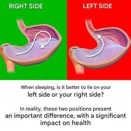
Here’s a detailed description of a medical illustration of the human stomach, which you could use as a reference for study or creating an image:
Anatomical Details of the Stomach
1. Location and Shape
- J-shaped, hollow muscular organ
- Located in the upper-left abdomen, beneath the diaphragm
- Connects the esophagus to the duodenum (first part of the small intestine)
2. Key Regions
- Cardia: where food enters from the esophagus
- Fundus: the upper rounded portion that stores undigested food and gases
- Body (Corpus): the main central region where mixing and digestion occur
- Antrum (Pyloric Antrum): lower portion that grinds food and regulates emptying
- Pylorus: narrow passage connecting to the duodenum, contains the pyloric sphincter
3. Layers of the Stomach Wall (from inside out)
- Mucosa: inner lining that secretes gastric juices and mucus
- Submucosa: connective tissue with blood vessels and nerves
- Muscularis externa: three layers of muscle (longitudinal, circular, oblique) for churning
- Serosa: outer protective layer
4. Internal Features
- Rugae: folds in the mucosa that allow the stomach to expand
- Gastric glands: produce acid (HCl) and digestive enzymes like pepsin
- Blood supply: mainly from branches of the celiac artery
- Innervation: parasympathetic (vagus nerve) and sympathetic fibers
5. Function Highlight
- Mechanical digestion: churning and mixing food
- Chemical digestion: secretes acid and enzymes to break down proteins
- Temporary storage before food passes to the small intestine
If you like, I can make a labeled, step-by-step visual diagram description that’s accurate enough for a student-level medical illustration — essentially a “ready-to-draw” guide.
Do you want me to do that?
