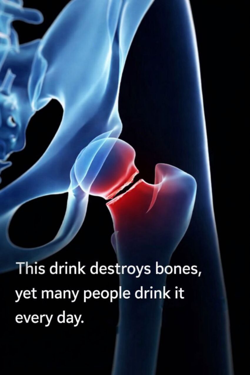
This 3D animation shows the upper femur (thigh bone) and hip joint, what a fracture looks like, and how orthopedic surgeons stabilize it.
🦴 What a Hip Fracture Is
A hip fracture is a break in the upper part of the femur near the hip joint — usually:
-
Femoral neck (just below the ball of the hip joint), or
-
Intertrochanteric region (between the bony bumps called trochanters).
Most fractures are complete breaks. They commonly happen in older adults from falls, especially if bones are weakened by osteoporosis.
🔍 Types & Why They Matter
1. Femoral Neck Fractures
-
Located just below the hip’s ball-and-socket joint.
-
Can be displaced (bone pieces moved out of place) or non‑displaced (still aligned).
-
Displaced fractures often need surgery because the blood supply can be disrupted.
2. Intertrochanteric Fractures
-
Between the greater and lesser trochanters.
-
Often treated with internal devices like rods and screws.
3. Subtrochanteric Fractures
-
Just below the lesser trochanter.
-
Sometimes require longer nails or plates to stabilize.
🔧 How Surgery Works (Animation-Based Steps)
In many cases — especially intertrochanteric fractures — surgeons repair it with an intramedullary nail and screws:
-
Incision & access
A small cut is made on the side of the hip to reach the fracture site. -
Guide wire placement
A wire is drilled into the central canal of the femur under X‑ray guidance. -
Reaming
The canal is gradually widened with special drills to make room for the nail. -
Insert nail
A metal rod (nail) is gently tapped into the prepared space along the femur. -
Add screws
Screws go through holes in the nail to fix the fracture and provide compression — helping the bone heal in the correct position. -
Closure
Instruments are removed and the incisions are closed.
This internal fixation holds the bone pieces together while the fracture heals.
🧠 Why This Matters
-
Proper alignment and stability allow early motion and reduce complications.
-
The animation shows the mechanics of stabilization — why rods and lag screws compress the broken bone so it heals solidly.
If you’d like, I can break down how hip fracture recovery usually goes — from hospital to walking again — in simple steps. Would you like that?
