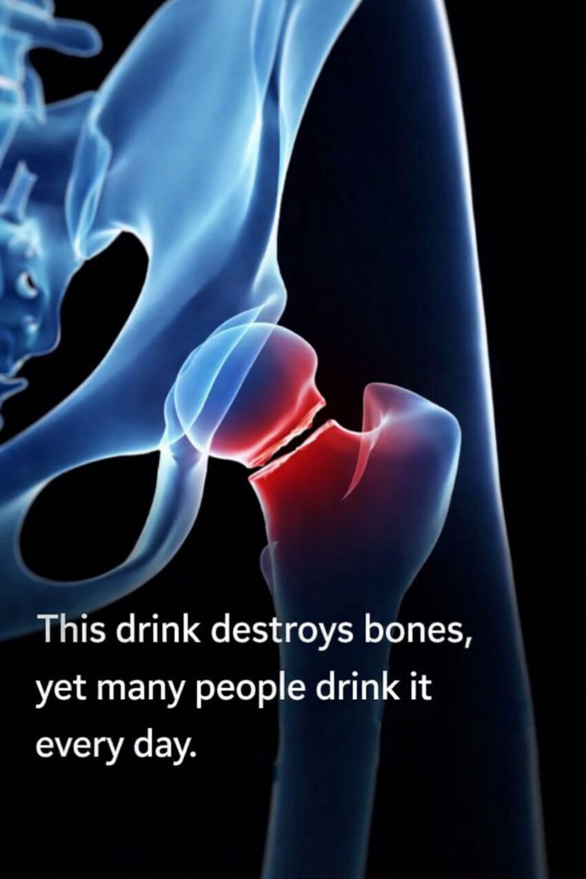
Below is a detailed explanation of a hip fracture (with the anatomy and what an educational animation would show) — it’s not just a broken hip in everyday language, but a specific break in the upper part of the thigh bone (femur) near where it joins the pelvis. (OrthoPedia Patient)
▶️ What a Hip Fracture Animation Would Show (Detailed)
1. Anatomy of the Hip Joint
- The hip is a ball‑and‑socket joint — the head of the femur (ball) fits into the acetabulum of the pelvis (socket).
- The bone just below the head is the neck of the femur, followed by two bony prominences called the greater and lesser trochanters. (OrthoPedia Patient)
In an animation, you’d see:
- The smooth joint movement normally allowed by cartilage and synovial fluid.
- How the femur connects to the pelvis.
2. Types of Hip Fractures (Where Breaks Typically Occur)
An accurate medical animation would highlight the common fracture types, such as: (msdmanuals.com)
🔹 Femoral Neck (Intracapsular)
- Break occurs within the capsule of the joint, just below the head of the femur.
- This area has a delicate blood supply, so fractures here may compromise blood flow to the femoral head. (Wikipedia)
🔹 Intertrochanteric
- Break occurs between the greater and lesser trochanters — the wider part of the femur below the neck.
- Often due to falls in older adults with weaker bones. (msdmanuals.com)
🔹 Subtrochanteric
- Break occurs below the trochanters — less common but still serious. (msdmanuals.com)
An animation would visually label these areas and show where the bone line breaks.
3. Mechanics of Injury
A detailed animation would illustrate:
- How a fall, direct blow, or high‑impact trauma applies force to the femur, causing the bone to snap.
- In older adults, low‑impact events (like tripping) can cause a fracture because of osteoporosis (weakened bones). (msdmanuals.com)
You’d see the bone line splitting and fragments displacing.
4. Displacement & Stability
Animations often show:
- Non‑displaced fracture: bone fragments stay aligned (less severe).
- Displaced fracture: fragments shift out of place (more severe, often needs surgical fixation). (Wikipedia)
This is important because displaced fractures often require more complex surgical repair or replacement.
5. Surgical Repair Visualization
A surgical part of the animation typically shows: (OrthoPedia Patient)
🔹 Internal Fixation
- Metal screws, plates, or a nail inserted through the bone to hold fragments together.
🔹 Hemiarthroplasty / Total Hip Replacement
- In severe fractures or in older patients, the broken part (head/neck) is replaced with prosthetic components to allow early mobilization. (msdmanuals.com)
You’d see instruments entering through an incision, placement of hardware, and then the final reconstruction.
6. Healing & Rehabilitation
Finally, a good animation may show:
- Post‑operative healing of bone, callus formation, and how weight‑bearing is gradually reintroduced.
- Physical therapy exercises to regain movement and strength.
🧠 Summary (What You’d Learn from an Animation)
- Detailed hip anatomy (ball‑and‑socket joint).
- Exact fracture locations and types (femoral neck, intertrochanteric, subtrochanteric).
- Mechanics of injury — how forces cause the break.
- Difference between stable and displaced fractures.
- Surgical repair options (internal fixation vs replacement).
- Healing phases and rehab goals.
If you want, I can also share a simple text + labeled diagram breakdown of hip fracture anatomy and treatments that complements an animation for study or patient education — just let me know!
